
Magnetic Resonance Imaging Department at the Dighomi Branch
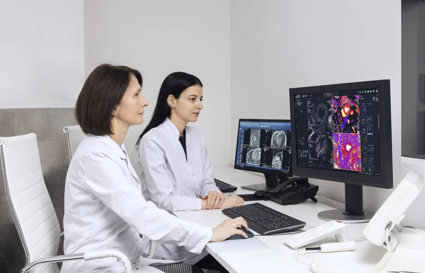
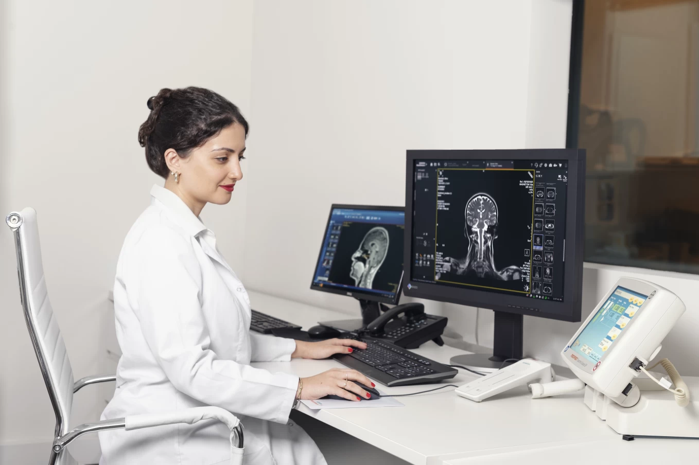
Head of the department - Mariam Kutateladze.
The Magnetic Resonance Imaging Department is equipped with two state-of-the-art magnetic resonance imaging systems from Siemens: Magnetom Lumina (3T strength), which is distinguished by:
- The latest generation of software, enabling detailed evaluation of structural changes in various pathologies.
- The capability to examine patients with high body weight.
- A wide tunnel ensuring comfort for claustrophobic patients during the examination.
- If necessary, the examination can be conducted under sedation. The anesthesiology department provides sedation for both adult and pediatric patients.
The Magnetic Resonance Imaging Department performs:
- Magnetic Resonance Imaging (MRI) of the Brain
The use of submillimeter slices significantly increases the likelihood of detecting any fine-fiber damage. - Spectroscopic study of pathological areas in the brain
Allows assessment of their metabolic level. - Detection of ischemic brain injury in the acute stage, and differentiation of tumor formation characteristics using diffusion-weighted imaging.
- Magnetic Resonance Angiography (MRA) of the brain and neck major blood vessels
This method is a non-invasive alternative to conventional X-ray contrast examination. - MRI of abdominal organs:
Liver, spleen, pancreas, adrenal glands, and kidneys; non-contrast Magnetic Resonance Cholangiopancreatography (MRCP) and Magnetic Resonance Angiography (MRA); MR enterography. - Magnetic Resonance Diagnostics of pelvic organ diseases, including multiparametric studies of the prostate and bladder.
- Magnetic Resonance Imaging of musculoskeletal structural disorders.
- MRI of pediatric patients.