Ultrasound Diagnostics Department at the Vera Branch
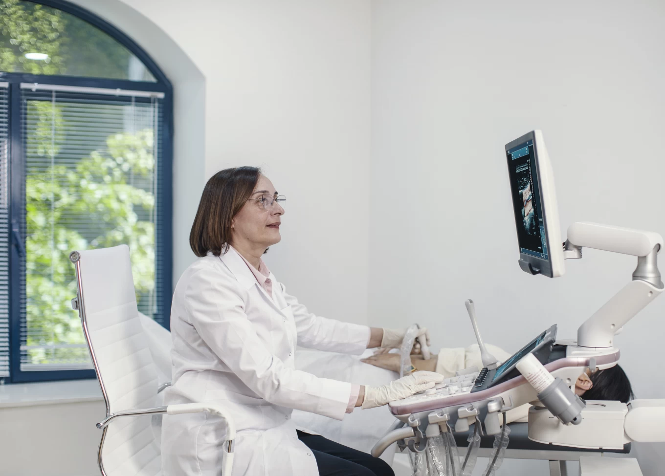
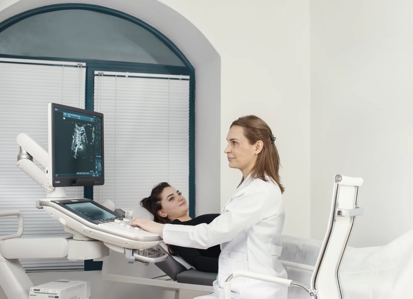
In the department of ultrasound diagnostics examinations are carried out using Canon ultrasound diagnostic devices of premium class: Aplio I 700, Aplio a.
The devices are distinguished by the latest software, have a large number of automated modern modes, high quality of visualization. The said enables high-quality scanning, visualization and detail to help you make an accurate diagnosis.
The following is available in the department:
- Abdominal ultrasound (liver, gallbladder, pancreas, spleen, liver gate dopplerography)
- Ultrasound of the urinary system (kidneys, adrenal glands, prostate, bladder)
- Gynaecological transabdominal and transvaginal ultrasound (uterus, ovaries), follicle monitoring
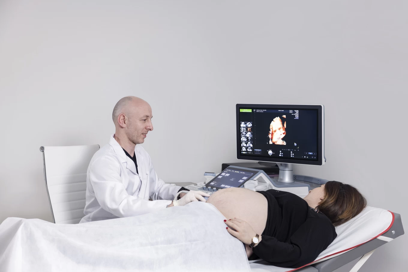
- Ultrasound examination of the fetus (first, second and third trimester of pregnancy)
- Thyroid gland ultrasound examination
- Mammary gland ultrasound examination
- Soft tissue ultrasound examination
- Ultrasound examination of regional lymph nodes
- Duplex scanning of blood vessels (extracranial, transcranial, upper and lower extremities, abdominal aorta, renal blood vessels)
- Ultrasound of joints
- Ultrasound examination of the hip joints of infants
- Cardiac echocardiography (for pregnant women after 24 weeks)
The main advantage of the device is:
ibeam ultrasound technology - provides high-quality, detailed images at any depth. The function of translucent glass ‘’Shadow Glass” makes it possible to examine the details with high quality.
The IDMS software maintains the image clarity at any depth.
Aplio sensors are highly sensitive. They can cover both deep and surface structures, and have the ability to visualize twice as much space as conventional sensors.
The SMI function makes it possible to assess the flow of blood in the micro-blood vessels.
Dr. Dan Skyby, Director of the Ultrasound Department at Toshiba America Medical Systems, said: ''We have developed technologies that are fully focused on safety, the comfort of a patient, ease of examination, and that gives a doctor the ability to be confident in his diagnosis.''
Obstetrical and gynaecological examinations in the Department of Ultrasound Diagnostics are carried out with the help of the latest generation device - GE Voluson E10 - BT19, the leader in obstetrical and gynaecological examinations:
- Best 3D/4D fetal image
- The new fetalHQ program evaluates the size, shape, and contraction of a fetal heart chambers
- Evaluation of fetal brain structures is much more effective and simple
- With an even better 2D, 3D/4D image quality, it is possible to more accurately identify pathological structural changes and abnormalities of the uterus and its appendages.
The special program Radiantflow quickly and easily visualizes blood circulation, including low-speed blood flows. High sensitivity combined elastography method with both dynamic visual elastography and SWE quantitative analysis and wave propagation maps.