
Department of Magnetic Resonance Imaging at the Vera Branch
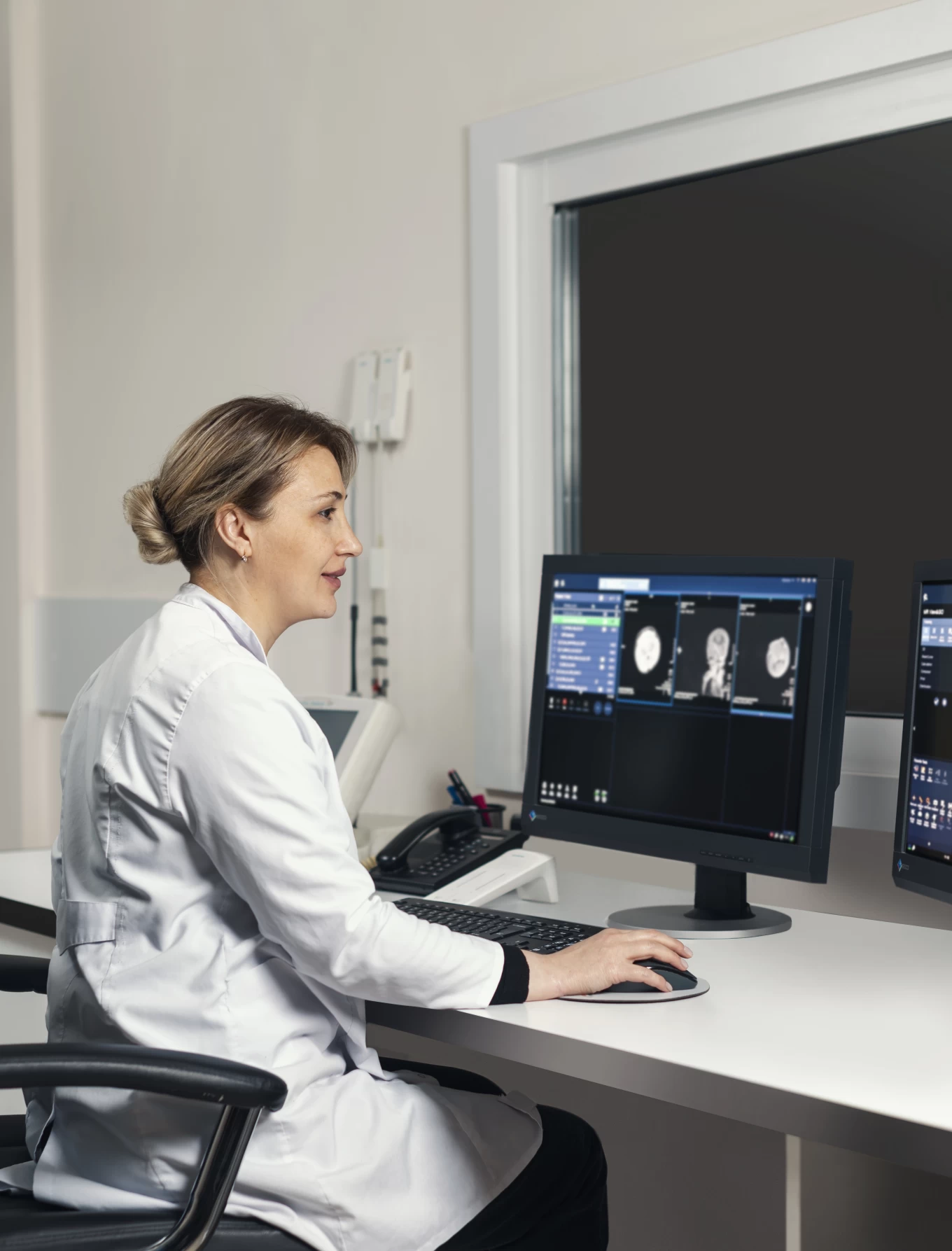
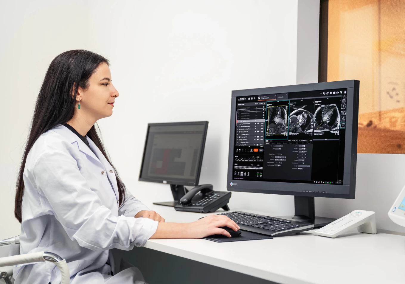
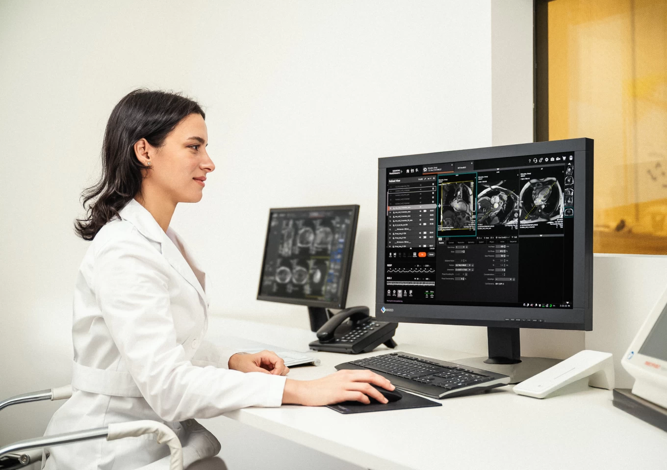
The Department of Magnetic Resonance Imaging is equipped with the latest magnetic resonance imaging from SIEMENS - Magnetom Vida (3T power), which is prominent with:
- The latest generation of software provides a detailed assessment of structural changes during various pathologies
- The ability to conduct examinations among patients with high weight
- The wide tunnel provides comfort for patients with claustrophobia during examination
- If necessary, it is possible to conduct a sedation examination, the anaesthesia service provides sedation for both adults and children
The following examinations are performed in the Department of Magnetic Resonance Imaging:
- Magnetic Resonance of the brain.
- The use of submillimetre sections highly increases the probability of detecting any small lesions
- Spectroscopic examination of the pathological area detected in the brain allows its assessment at the metabolic level
- Detection of ischemic brain damage in the acute stage and differentiation of the nature of tumour formations are possible using diffusion-weighted images
- Magnetic resonance angiography of the main blood vessels of the brain and neck - this method is a non-invasive alternative to conventional X-ray imaging
- Examination of the abdominal organs: liver, spleen, pancreas, adrenal glands and kidneys, non-contrast magnetic resonance cholangiopancreatography, magnetic resonance angiography, MR enterography
- Magnetic resonance diagnostics of diseases of the pelvic organs, including prostatitis, and multiparametric examinations of the bladder
- Magnetic resonance study of structural disorders of the musculoskeletal system
- Magnetic resonance examination of the heart using functional, morphological and late contrast enhancement modes, which gives important additional information in the diagnostics of inflammatory, infiltration, ischemic heart diseases and cardiomyopathy
- Magnetic resonance examination of children