
Department of Nuclear Medicine
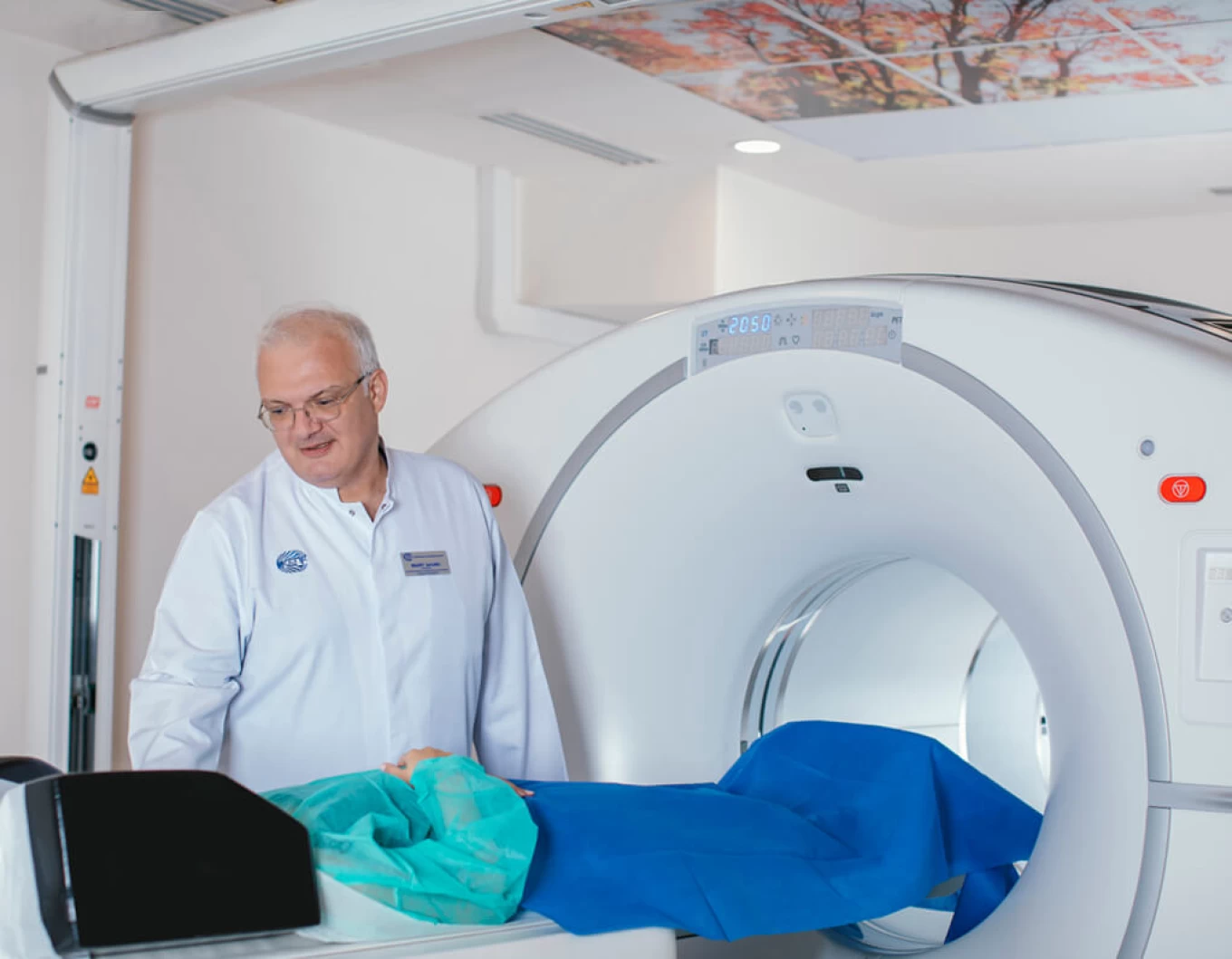

Head of the Department - Michael Baramia
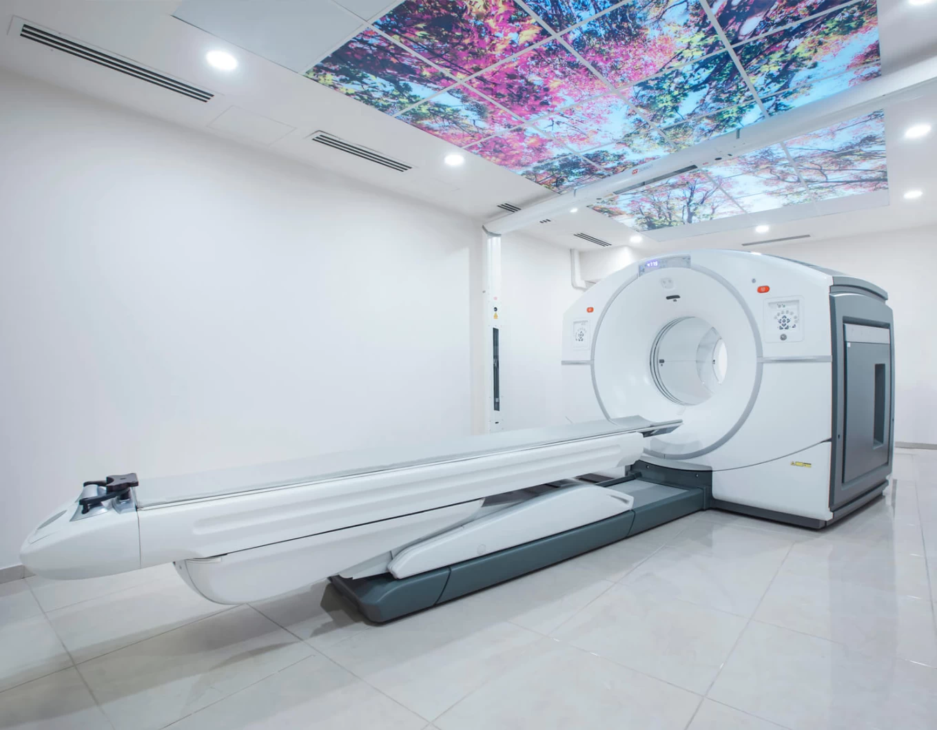
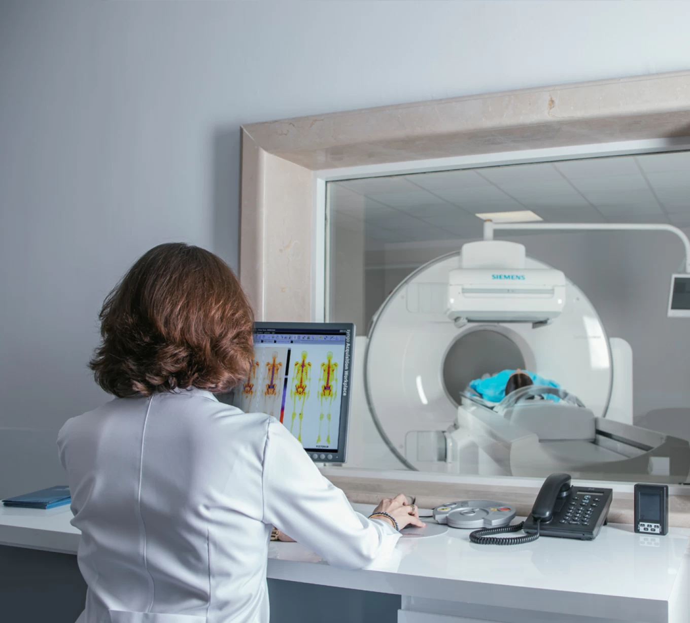
With the development of Hybrid Positron Emission Tomography / Computed Tomography (PET/CT), it became possible to combine metabolic and anatomical data. The introduction of this technology has had a significant impact on various medical disciplines, including neurology and cardiology, but it has particularly revolutionized the field of oncology. Today, Positron Emission Tomography (PET/CT) is widely used worldwide in the management of oncological diseases. It provides the ability to determine the precise anatomical localization of metabolic activity, making PET/CT indispensable in the staging, restaging, and evaluation of treatment responses in oncological diseases. In approximately 30% of cases, PET/CT alters the treatment strategy for oncology patients.
The principle of Positron Emission Tomography (PET) is based on the enhanced metabolism of glucose by tumor cells and tissues compared to healthy tissues. Glucose is labeled with a radioactive isotope of fluorine (F18, which has a half-life of 110 minutes), which emits positrons during radioactive decay. The gamma rays produced from the interaction of positrons with electrons are detected by the scanner's detectors, providing information about the metabolic activity of different areas of the body. This helps in the detection of tumor cells and tissues. For this reason, PET scans have found widespread use in oncology. Today, F18-fluorodeoxyglucose (FDG) is the most commonly used radiopharmaceutical for PET imaging worldwide.
Positron Emission Tomography (PET/CT) Indications in Nuclear Medicine:
-
Determination of the extent of cancer spread (staging/restaging): PET/CT is crucial in assessing how far a cancer has spread, helping doctors determine the stage of the disease and evaluate whether it has advanced or spread to other areas of the body.
-
Detection of recurrence and/or continuous growth: PET/CT can be used when clinical and anatomical data are negative, especially in cases where cancer markers are elevated, helping detect recurrence or further progression of the disease.
-
Detection of the primary lesion in metastatic and/or paraneoplastic syndrome: In cases of metastatic cancer or paraneoplastic syndromes, PET/CT helps identify the original tumor source, which may otherwise be difficult to locate with conventional methods.
-
Differentiation between benign and malignant tumors: PET/CT helps distinguish between benign (non-cancerous) and malignant (cancerous) tumors, which is vital for proper diagnosis and treatment planning.
-
Assessment of treatment effectiveness (chemotherapy, radiation therapy, combined therapies): PET/CT is used to evaluate the effectiveness of various treatment modalities like chemotherapy and radiation therapy, by showing how the cancer responds to treatment.
-
Selecting the optimal location for biopsy: PET/CT can help guide doctors in selecting the most suitable site for biopsy, ensuring that tissue samples come from areas with the highest likelihood of containing cancer cells.
-
Planning for surgical treatment: PET/CT is used to plan surgery by accurately identifying the extent of the tumor, its location, and its relation to nearby structures, which is essential for successful surgical intervention.
-
Planning for radiation therapy: PET/CT plays a crucial role in radiation therapy planning, helping to target tumors precisely and avoid irradiating healthy tissues, improving the overall effectiveness of the treatment.
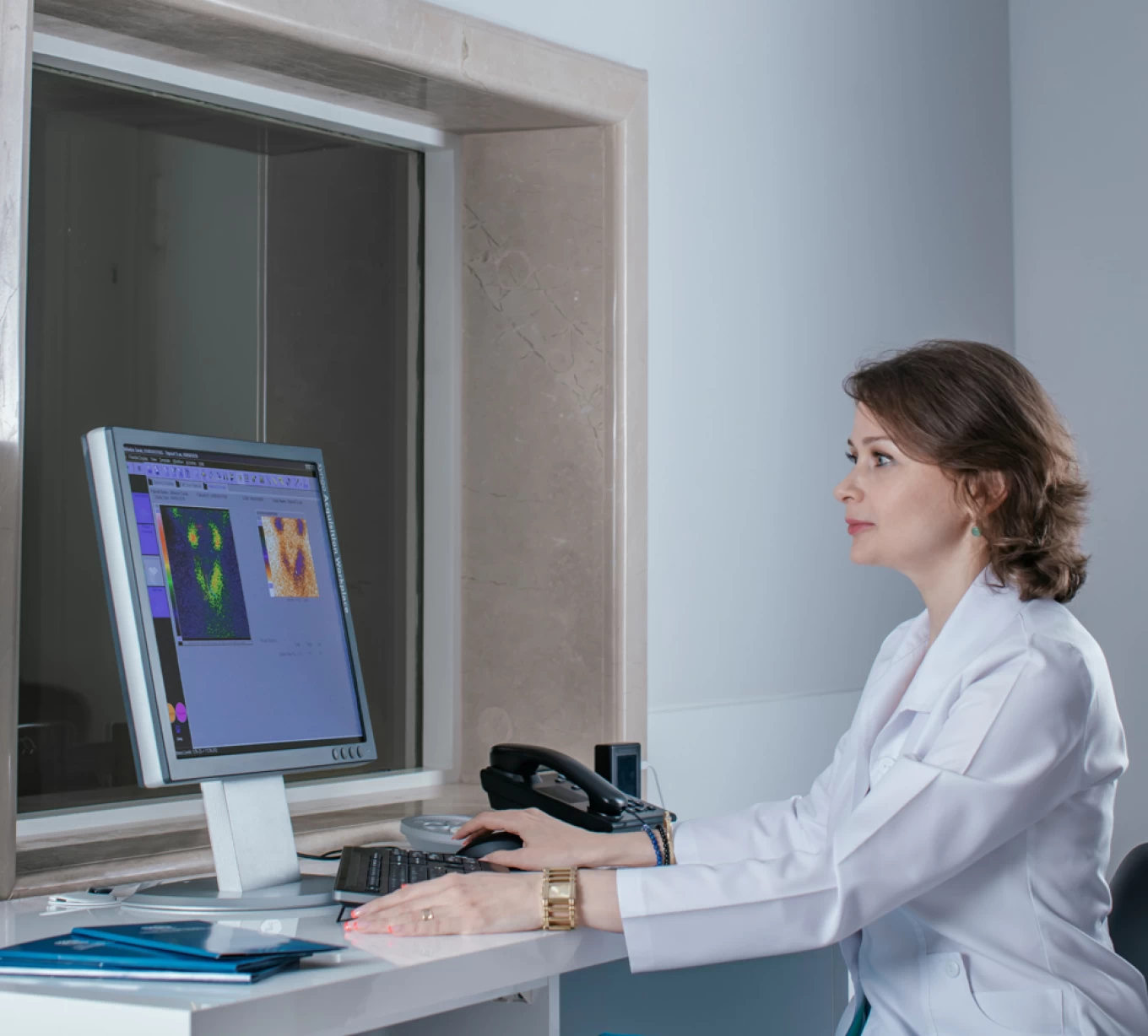
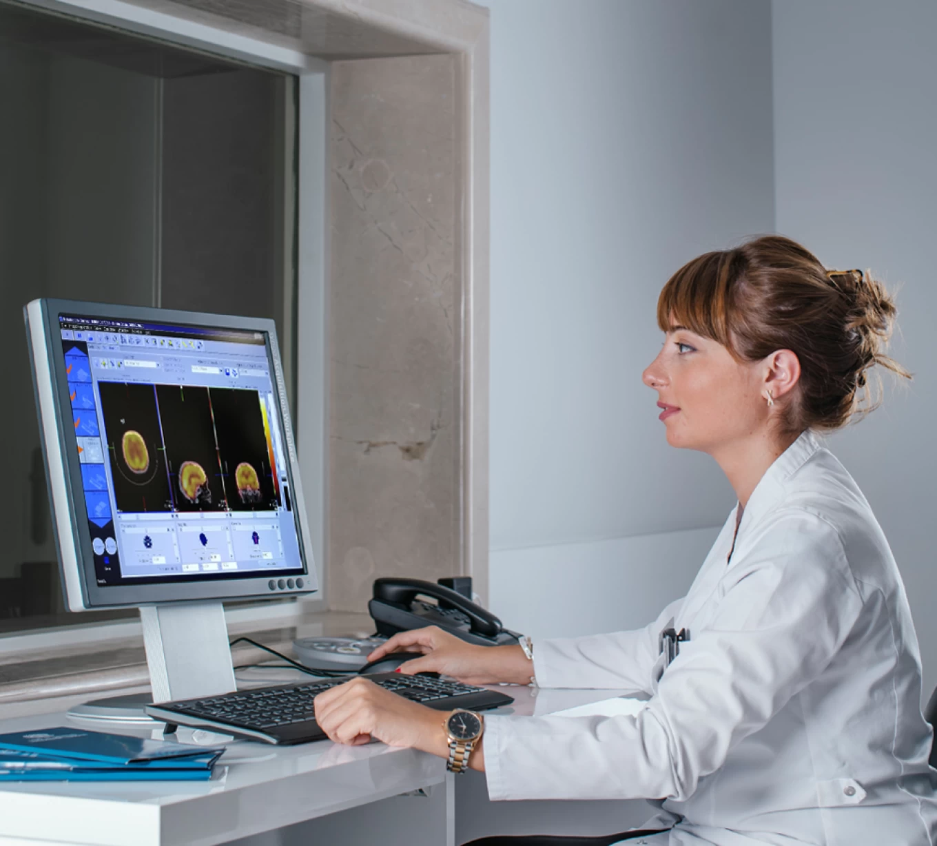
Head of the Radionuclide Diagnostics Department - Tatia Aleksishvili
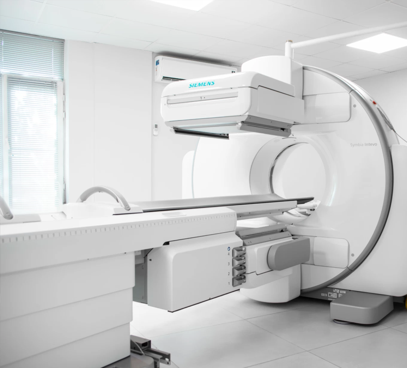
The Nuclear Medicine Department at Todua Clinic is equipped with two state-of-the-art devices from SIEMENS, namely the Symbia Intevo SPECT/CT and the Symbia Evo single-photon emission tomography (SPECT) systems.
These advanced technologies enable us to perform all modern diagnostic procedures at an international level with much greater accuracy. The equipment makes diagnostic procedures easier for patients as it allows for faster scans with minimal radiation exposure.
Radionuclide imaging plays a significant and irreplaceable role in assessing the function of various organs.
Radionuclide Diagnostics Department performs:
Radionuclide Diagnostics Department performs:
- Bone Scintigraphy in oncology patients (for detecting metastatic damage) and in traumatology patients (for diagnosing bone osteomyelitis and prosthesis loosening)
- Thyroid Scintigraphy (for determining the cause of thyrotoxicosis; also used to assess the functional condition of thyroid nodules, to identify hyper- and/or hypofixation areas)
- Parathyroid Scintigraphy (for detecting parathyroid adenoma)
- Dynamic and Cortical Nephroscintigraphy (for assessing kidney functional condition and GFR; for detecting congenital kidney anomalies, renal artery stenosis, and renovascular hypertension; for diagnosing kidney trauma, scarring, urinary tract infections, and bladder-ureteral reflux; it is virtually indispensable for diagnosing obstructive kidney diseases)
- Gastric Scintigraphy (for evaluating gastric motility and evacuation function – also known as gastric emptying scan)
- Meckel's Diverticulum Scintigraphy (the most reliable method for detecting Meckel's diverticulum, the most common congenital disorder of the gastrointestinal tract, and determining the cause of melena in young children)
- Bone Scintigraphy in Traumatology Patients
- Myocardial Scintigraphy (for diagnosing myocardial blood supply issues)
- Lymphatic System Scintigraphy (for evaluating lymphatic drainage)