Magnetic Resonance Imaging Department - MRI
Magnetic Resonance Imaging at Todua Clinic
Department Head - Ketevan Lashkhi
The Magnetic Resonance Imaging (MRI) Department is equipped with six MRI machines from the company SIEMENS:
-
- Magnetom Vida, Magnetom Skyra, Magnetom Skyra Fit, and Magnetom Lumina (3T strength)
- Magnetom Aera and Magnetom Avanto Fit (1.5T strength)
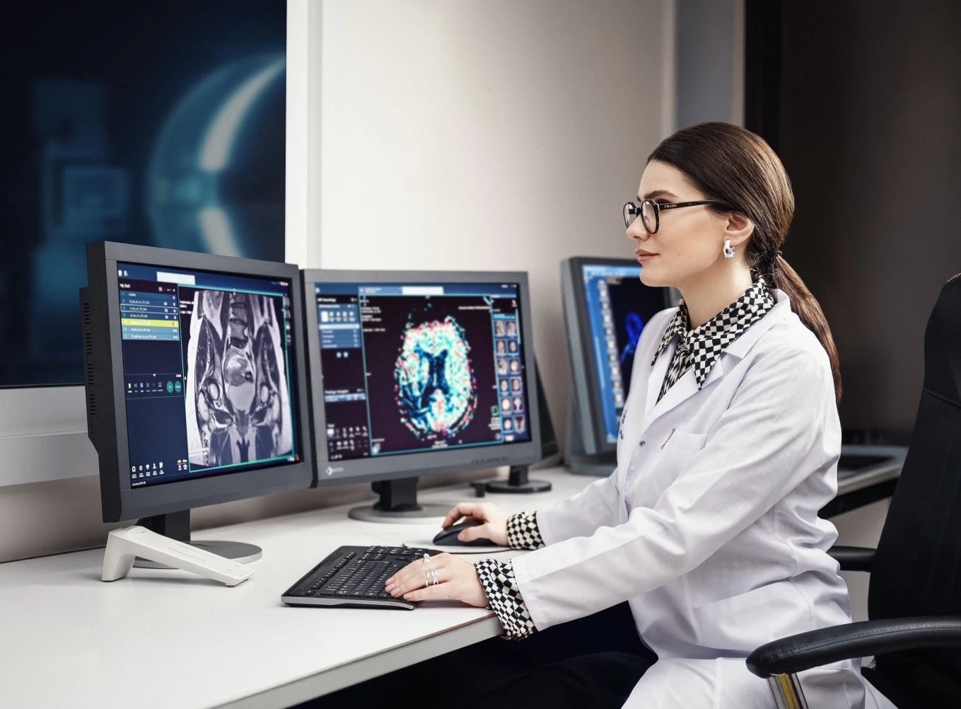
These MRI machines are distinguished by high image quality. They are equipped with comprehensive software support for various specialties, including neurological, angiological, cardiological, oncological, orthopedic, abdominal and pelvic, as well as pediatric imaging.
Our medical center's MRI machines are distinguished by:
- State-of-the-art software equipment, allowing for the evaluation of both structural and functional changes.
- Specialized pediatric software.
- The ability to conduct spectroscopy, tractography, and liver Liver Lab assessments.
- The widest and shortest tunnels in the region, equipped with Mood Light lighting to ensure comfort during the examination of claustrophobic patients.
- The 2021 SyngoVia work platform, featuring the latest 4G, Dot, Tim, and GO functions.
- The possibility of examining heavy-weight patients.
- The ability to conduct full-body and mammography scans.
If necessary, examinations can be performed under sedation. The anesthesiology department ensures sedation for both adult and pediatric patients during the procedure.
Whole-body examinations are conducted using both non-contrast and contrast-enhanced imaging. In the case of detecting a pathological focus, additional local area imaging is performed.
MRI Examination
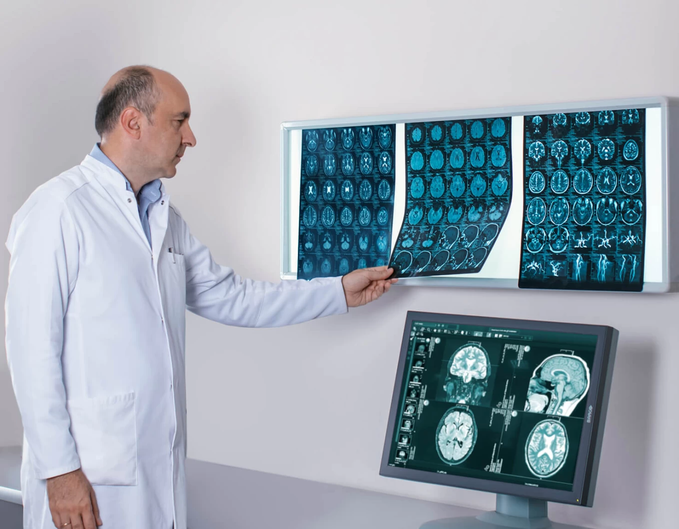
- Structural brain imaging, including submillimeter slice imaging, which significantly increases the likelihood of detecting any fine-grained damage.
- Magnetic Resonance Angiography (MRA) of the brain and cervical major blood vessels – this method is a non-invasive alternative to conventional X-ray contrast imaging.
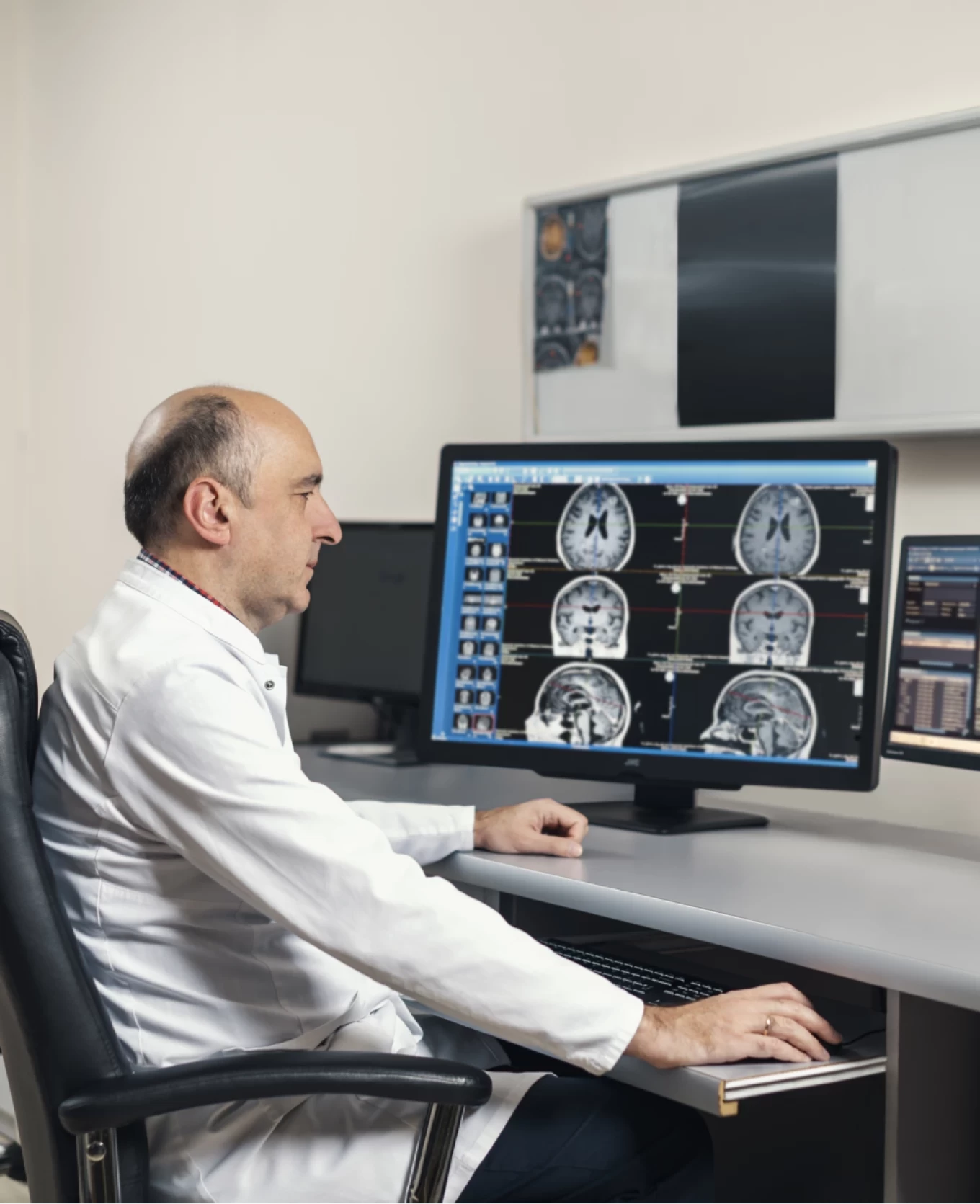
- Functional brain imaging for the study of higher neural functions, localization of cortical centers, and examination of speech, movement, vision, and memory. These methods assist in pre-surgical evaluations to avoid damage to significant cortical areas during surgery, as well as enabling early diagnosis of cognitive function impairments.
- White matter tractography – the study of the integrity, direction, and connections of white matter fibers. Identifying the relationship of pathological foci to functionally significant tracts.
- Disease prognosis based on the study of white matter damage, including brain perfusion and diffusion imaging, detection of ischemic damage at the acute stage, and differentiation of the nature of neoplastic formations.
- Fundamental research in the field of neuroscience – functional memory studies and brain tractography research.
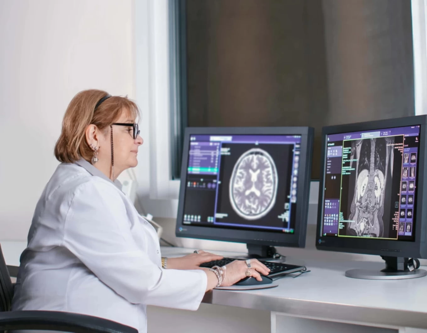
- Examination of abdominal cavity organs: liver, spleen, pancreas, adrenal glands, and kidneys; non-contrast Magnetic Resonance Cholangiopancreatography (MRCP); dynamic kidney imaging and Magnetic Resonance Angiography (MRA).
- Magnetic resonance diagnostics of pelvic organ diseases.
- Magnetic Resonance Imaging (MRI) of musculoskeletal system structural and functional disorders.
- MRI for pediatric patients.
It is noteworthy that the direction of Magnetic Resonance Imaging (MRI) was established for the first time in the region in 1994 at our medical center by Academician Fridon Todua.
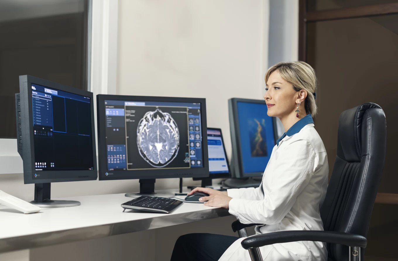
Doctors with many years of experience working in the department constantly participate in and lead sections at congresses of the European and American Radiology Associations, oversee international directions of radiological clinical research, and regularly publish articles in various international journals. The high professionalism of each of them and their thoughtful attitude toward patients should be emphasized.
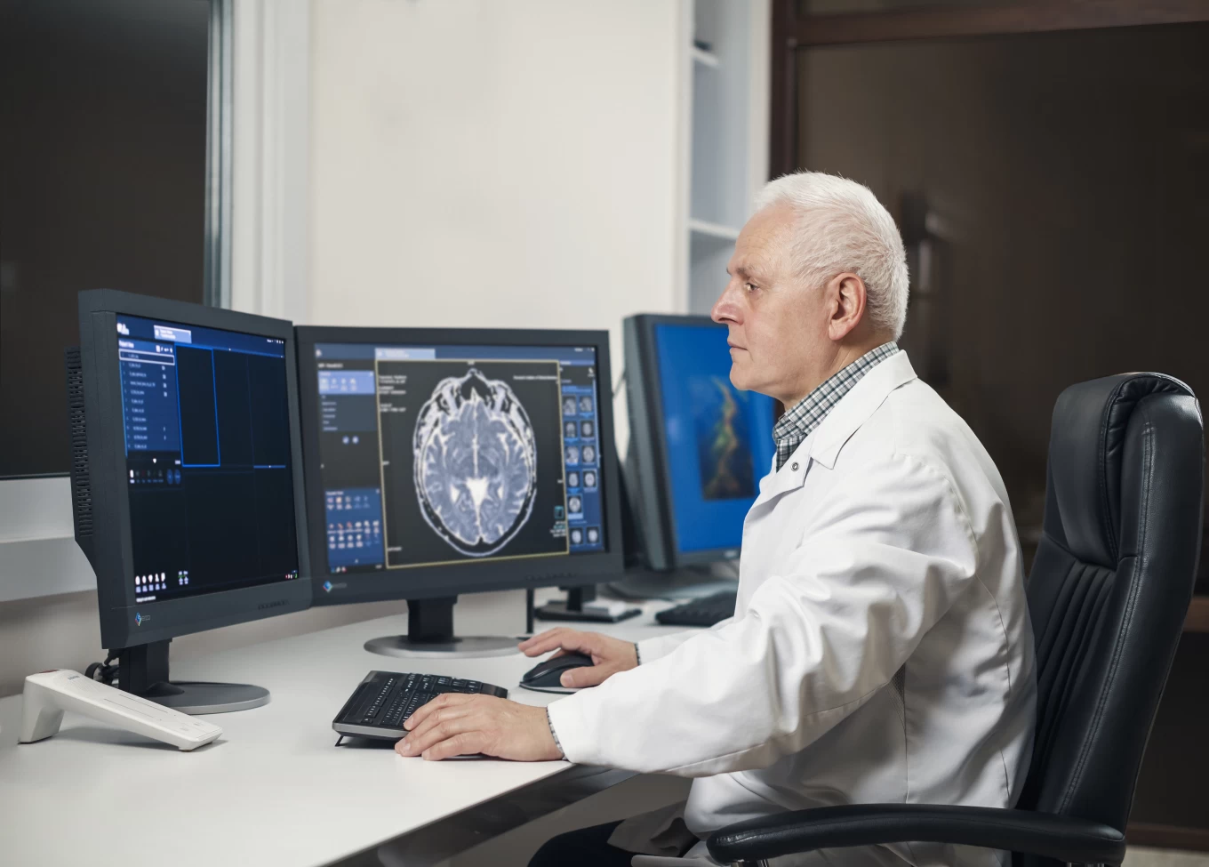
MRI Scanning
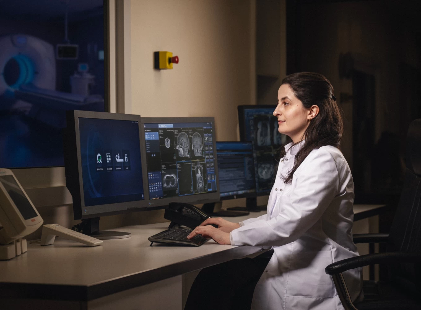
MRI scanning, also known as Magnetic Resonance Imaging (MRI), is a non-invasive medical practice that uses strong magnetic fields and radio waves to create detailed images. MRI is widely used in healthcare to visualize the structure and function of organs and various tissues. Magnetic Resonance Imaging provides critical information, which is utilized for diagnosing and monitoring various medical conditions.
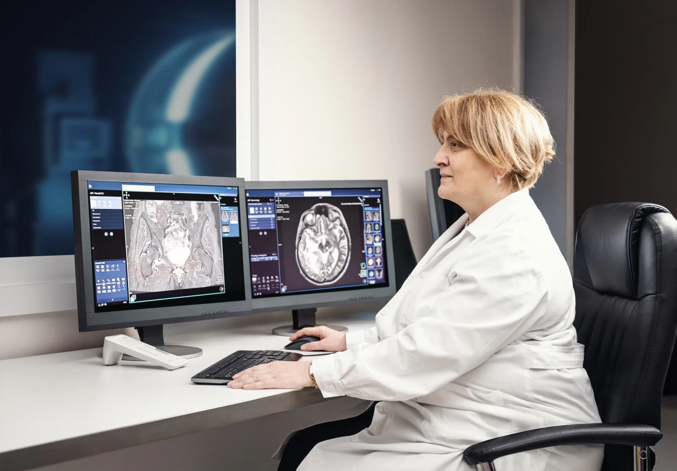
The MRI system by Siemens Healthineers is one of the best in the industry. The company’s MRI systems are designed to ensure high-quality imaging. In Todua Clinic, MRI scanning is performed at the highest level, and our team has many years of experience in this field.
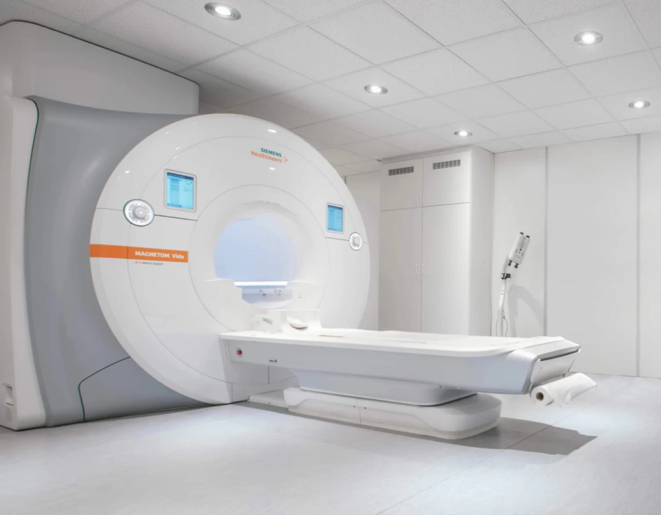
Magnetom Vida
Magnetom Vida is a magnetic resonance imaging (MRI) system with exceptional image quality and BioMatrix technology, which adapts to the unique physiological characteristics of each patient. MRI scanning can often be uncomfortable, but this machine helps reduce patient anxiety. It also enhances the image quality. The system’s powerful magnet and advanced imaging techniques allow for detailed images from various anatomical regions.
Magnetom Skyra
Another 3T MRI system, the Magnetom Skyra, is known for its versatility and capabilities. It is equipped with Tim 4G (Total Imaging Matrix) technology, which optimizes both image quality and acquisition speed.
Magnetom Skyra Fit
The Magnetom Skyra Fit is a compact version of the Magnetom Skyra 3T MRI system. This device makes MRI scanning much easier. Despite its compact size, it maintains high image quality and flexibility.
Magnetom Lumina
Introducing the Magnetom Lumina, equipped with a strong magnet and the latest imaging technologies. The system’s user-friendly interface and simplified workflows help ensure maximum comfort for patients while achieving highly effective MRI results.
Magnetom Aera
This is a 1.5T MRI system. It is equipped with Tim (Total Imaging Matrix) technology, which allows for high-quality imaging. (See detailed information about the system).
Magnetom Avanto Fit
Magnetom Avanto Fit is a compact 1.5 Tesla MRI system. It is equipped with Tim 4G technology.
Siemens Healthineers’ MRI systems combine advanced hardware, software, and patient-centered features to enhance diagnostic accuracy, simplify workflows, and ensure that patients are as comfortable as possible during their examinations. The diversity of 3T and 1.5T systems allows healthcare providers to choose the system that precisely meets their clinical and operational needs.
MRI Examination – Price and Quality
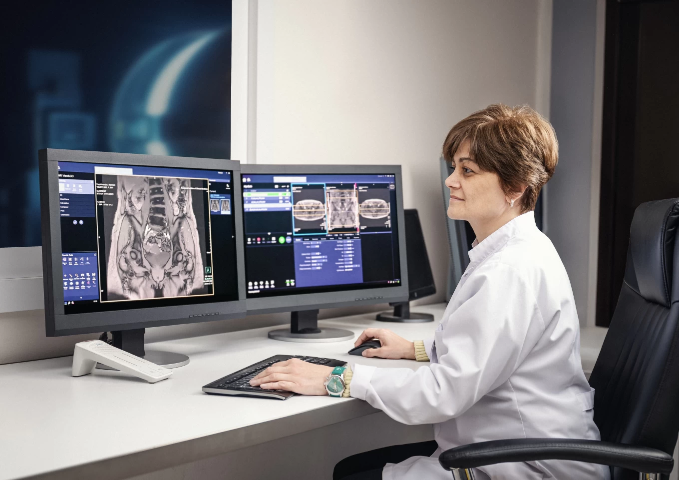
At Todua Clinic, the price and quality of MRI examinations are in perfect balance with each other. Patient care is our clinic's main priority. Our team of experienced professionals uses state-of-the-art technology and innovative methods to ensure that the treatment and diagnostic process is as effective as possible.
Of course, we strive to make all procedures accessible to our patients.
It is noteworthy that the first magnetic resonance imaging direction in the region was implemented in 1994 at our Medical Center by Academician Fridon Todua.
Experienced doctors working in the Department regularly participate and lead sections at congresses of the European and American Association of Radiology, lead directions in international radiological clinical trials, regularly publish articles in various international scientific publications. The high professionalism and worm attitude of each of them towards the patients is noteworthy as well.