
Laboratory of Pathomorphology
Head of the Department - Lali Tsivtsivadze, MD
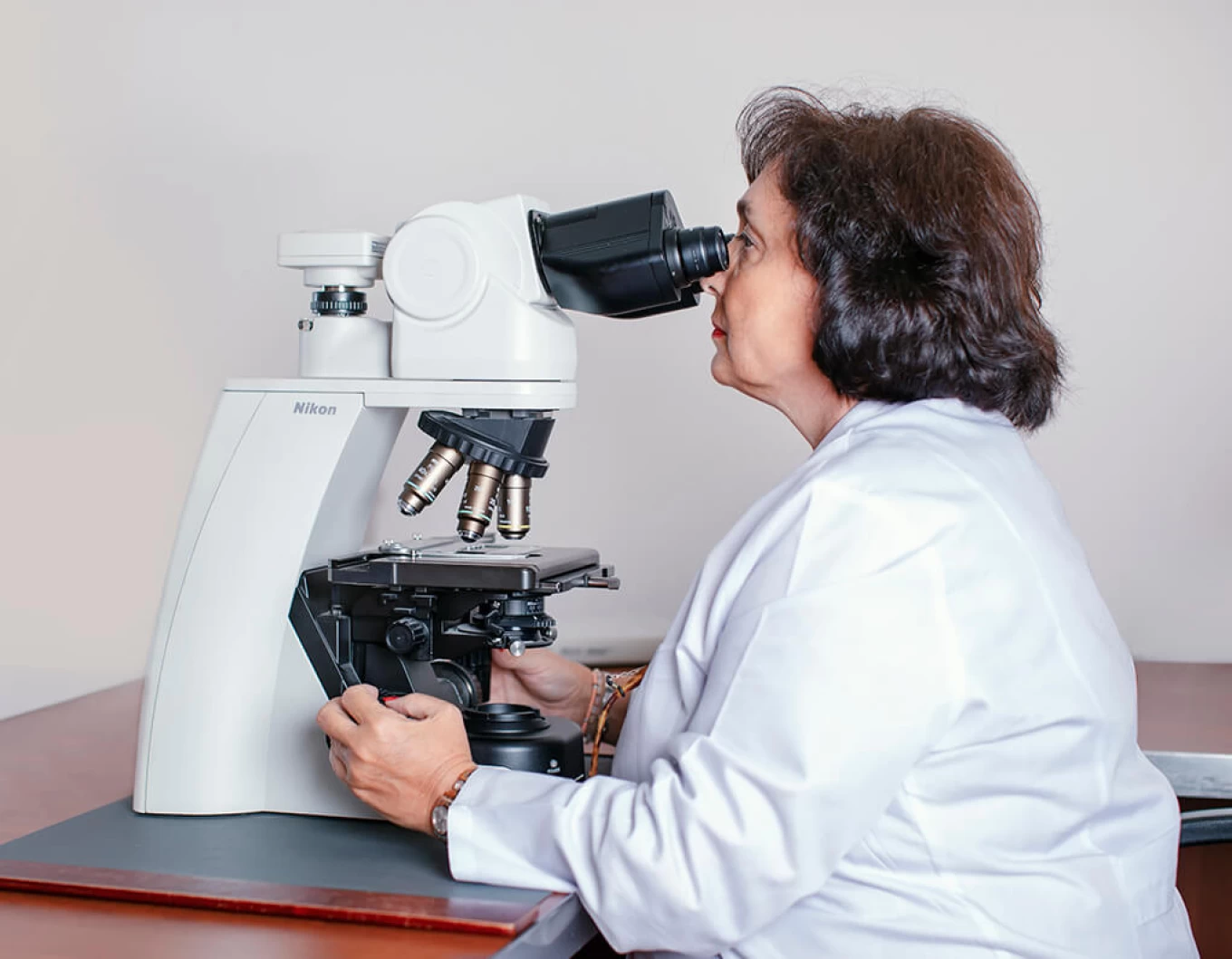
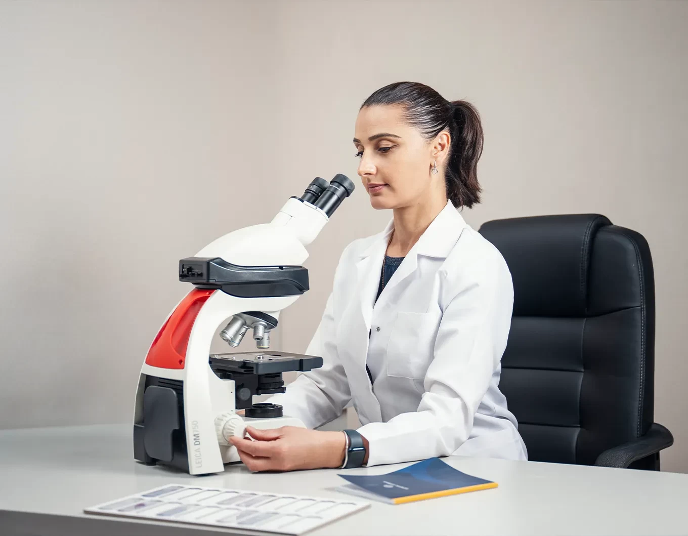
The Pathomorphology Laboratory is equipped with advanced LEICA equipment and digital microscopes: OLIMPUS BX 43, NICON ECLIPSECI, immunohistochemical analysis system LEICA BOND MAX, fully automated microtome – LEICA RM 2255 MICROTOME, tissue processor – LEICA ASP 6025, rapid diagnostic device – DIAPATH, automatic staining system – LEICA ST5020 MULTISTAINER. Additionally, the laboratory is equipped with a fluorescence microscope – AXIO IMAGER A2 FICH ANALYSIS for HER2/CEN analysis, and molecular testing – ALK status determination.
The Pathomorphology Department conducts the following:
- Examination of surgical, biopsy (including express biopsy), and cytological samples with general morphological, immunohistochemical, and in situ hybridization analyses.
- Immunohistochemical analysis is performed using the LEICA AUTOSTAINER BOND MAX system, utilizing a wide range of antibodies.
The mentioned method is used for:
- Confirmation of tumor histogenesis.
- Identification of the primary lesion.
- Determination of the degree of tumor anaplasia.
- Definition of immunophenotype in lymphoproliferative diseases.
- Determination of estrogen and progesterone receptors, as well as the oncogene CERBB/Her2/neu in cases of breast cancer.
- Assessment of tumor tissue sensitivity to hormonal and chemotherapeutic agents.
The main directions of the Pathomorphology Laboratory’s work include:
- Morphological diagnosis of pathological processes in various organs and systems during life.
- Clinicopathological analysis.
- Prognostic evaluation of processes.