
Department of Ultrasound Diagnostics
Head of the Department - Dudana Gachechiladze
Technological progress plays a significant role in the development of medicine. It is the new devices that provide the possibility of making an accurate diagnosis without unnecessary manipulations. One such technology is ultrasound equipment, which is used for safe and painless procedures.
Ultrasonic diagnostics, also known as ultrasonography, is a non-invasive method for detecting health problems. Ultrasound uses high-frequency sound waves to create visual images of internal organs. Ultrasound diagnostics allow professionals to examine soft tissues of internal organs in detail with the help of the images obtained and identify problematic areas.
Ultrasonic Diagnostics in Medicine
The use of ultrasound diagnostics in the medical field is broad and diverse. For example, the most well-known application of ultrasound is the monitoring of pregnant women. This procedure allows doctors to track fetal development, detect congenital anomalies or potential complications, determine the baby's position, and establish its birth date.
Moreover, ultrasonography is actively used in cardiology. Modern devices provide detailed images of various parts of the heart, valves, and blood vessels, which significantly contribute to the diagnostic process-helping assess damage to the cardiovascular system, based on which a treatment plan is prescribed.
Ultrasonic diagnostics is increasingly used for the visualization of muscles, tendons, and joints, as it allows for the detection of strains and other injuries. Additionally, ultrasonography is a non-invasive method for detecting diseases and complications in organs such as the liver, kidneys, pancreas, gallbladder, etc.
In addition to the aforementioned applications, ultrasound diagnostics is used in many other cases, such as obtaining images of blood vessels. Doppler ultrasound is a specialized technique that allows for the assessment of blood flow in arteries and veins.
Advantages
The main advantage of ultrasound diagnostics is the ability to make a reliable diagnosis, which is facilitated by the continuous development of the method and technological support. Innovations such as 3D and 4D ultrasound diagnostics contribute to the maximum accuracy in identifying health problems.
During ultrasound diagnostics, instead of radiation, only ultrasound waves are used, which are considered safe for patients, including pregnant women and infants. Additionally, this diagnostic method offers the possibility of reducing stress and physical pain for the patient, as the procedure does not require incisions or injections.
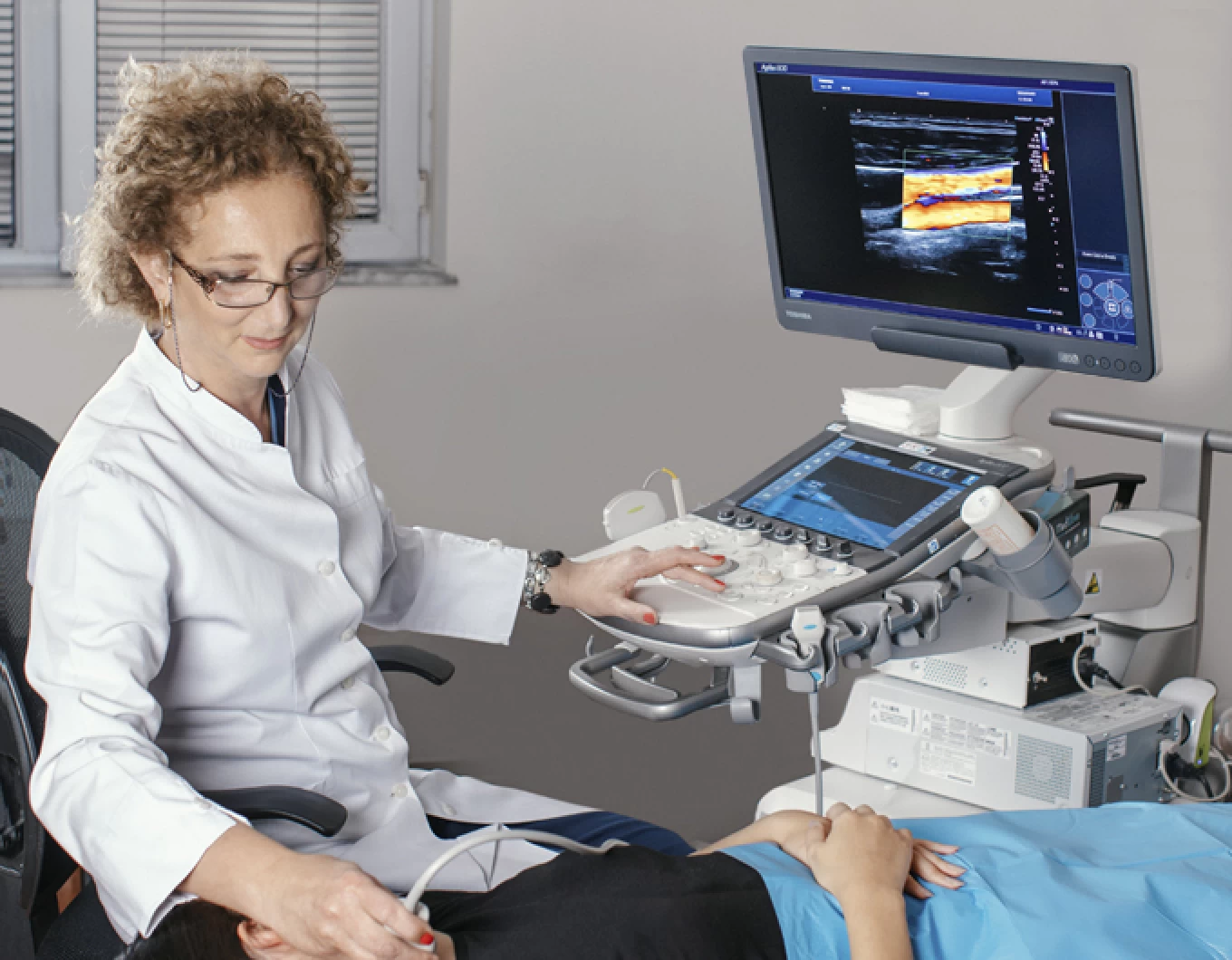
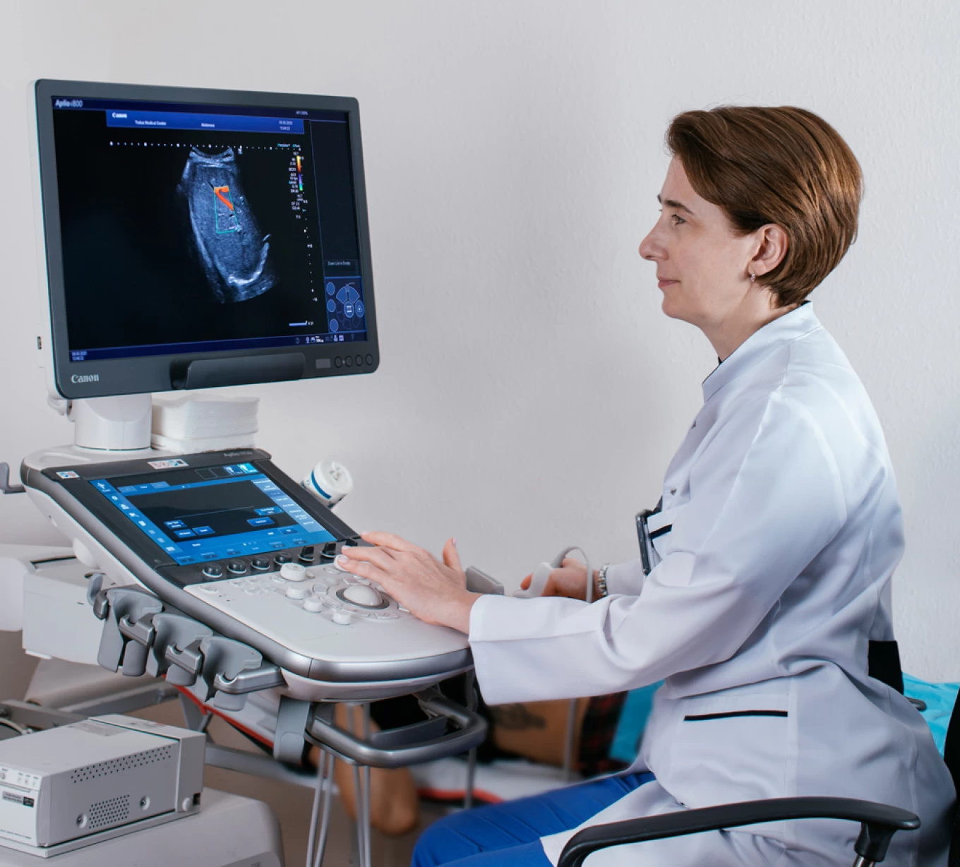
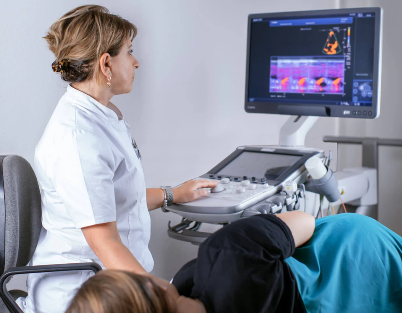
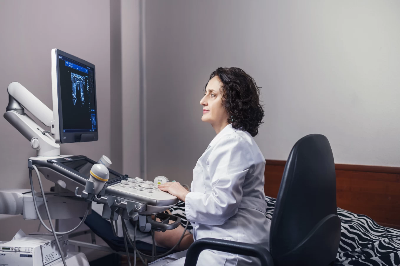
The department is equipped with premium-class, ergonomically designed ultrasound diagnostic devices, featuring the latest software, from the company Canon, including the Aplio i700, Aplio i800, Aplio i900, Aplio500, Aplio 300, Toshiba Aplio i800 Prism Edition, as well as the premium-class device from GE, the GE Voluson E10 - BT19.
Ultrasound Diagnostics at Todua Clinic
Todua Clinic is focused on offering innovative approaches, state-of-the-art equipment, and highly qualified services to its patients.
The Department Conducts:
- Hepatopancreatobiliary system
- Kidneys, adrenal glands, bladder, prostate
- Mammary glands, thyroid gland, and peripheral lymph nodes
- Pelvic organs
- Musculoskeletal system ultrasound examination
- Transthoracic echocardiography
- Extra- and intracranial blood vessels
- Abdominal cavity
- Duplex scanning of upper and lower limb blood vessels
- Early diagnosis of fetal developmental anomalies
- Elastographic assessment of the thyroid, mammary glands, abdominal cavity organs, soft tissue masses, and lymph nodes
- Quantitative examination of diffuse (fibrotic) changes in the liver using shear wave elastography (SWE) method
- Diagnostic puncture biopsy under ultrasound control for various pathologies
- Conducting therapeutic and diagnostic procedures
The Canon I Series is distinguished by exceptional crystal-clear images and high resolution. Unmatched speed and superior image quality ensure ideal visualization of even the smallest details.
The revolutionary iBeam architecture technology of Aplio and next-generation matrix transmitters allow for unprecedented high-quality images at practically any depth.
Through the iDMS (Intelligent Dynamic Microslice) program, image sharpness is maintained at any depth of examination, providing the highest resolution image quality that not only enables the most accurate diagnostics but also allows for precise performance of various interventional procedures (biopsy, puncture, drainage) under ultrasound control.